
Hip Replacement Surgery in Jaipur
Total & Partial Hip Arthroplasty by Dr. Hemendra Agrawal

Dr. Hemendra Agrawal
MBBS, MS (Orthopedics) · Senior Orthopedic Consultant, Narayana Hospital
Overview
What is Hip Replacement Surgery?
Hip Replacement Surgery, also known as Hip Arthroplasty, is a surgical procedure in which the damaged or diseased hip joint is replaced with an artificial prosthetic implant. The hip joint is a ball-and-socket joint where the head of the femur (thigh bone) articulates with the acetabulum (socket) of the pelvis. When arthritis, injury, or disease damages this joint, it can cause debilitating pain, stiffness, and loss of mobility that significantly impacts quality of life.
Dr. Hemendra Agrawal performs both total hip replacement (replacing both the ball and socket) and hemiarthroplasty (replacing only the femoral head, commonly done for hip fractures in elderly patients). He utilizes advanced bearing surfaces including ceramic-on-ceramic, ceramic-on-polyethylene, and oxidized zirconium-on-polyethylene, selecting the optimal combination based on each patient's age, activity level, and anatomy.
Using the direct anterior approach when appropriate, Dr. Agrawal performs hip replacement through a muscle-sparing technique that accesses the hip joint between muscles rather than cutting through them. This approach allows faster recovery, reduced risk of dislocation, less post-operative pain, and earlier return to normal activities compared to traditional posterior or lateral approaches.
Advanced uncemented (press-fit) implants with porous-coated surfaces encourage biological bone ingrowth, providing long-term fixation without cement. For elderly patients with poor bone quality, cemented fixation provides immediate stability. Dr. Agrawal's comprehensive approach, from pre-operative optimization through rehabilitation, ensures optimal outcomes for every patient.
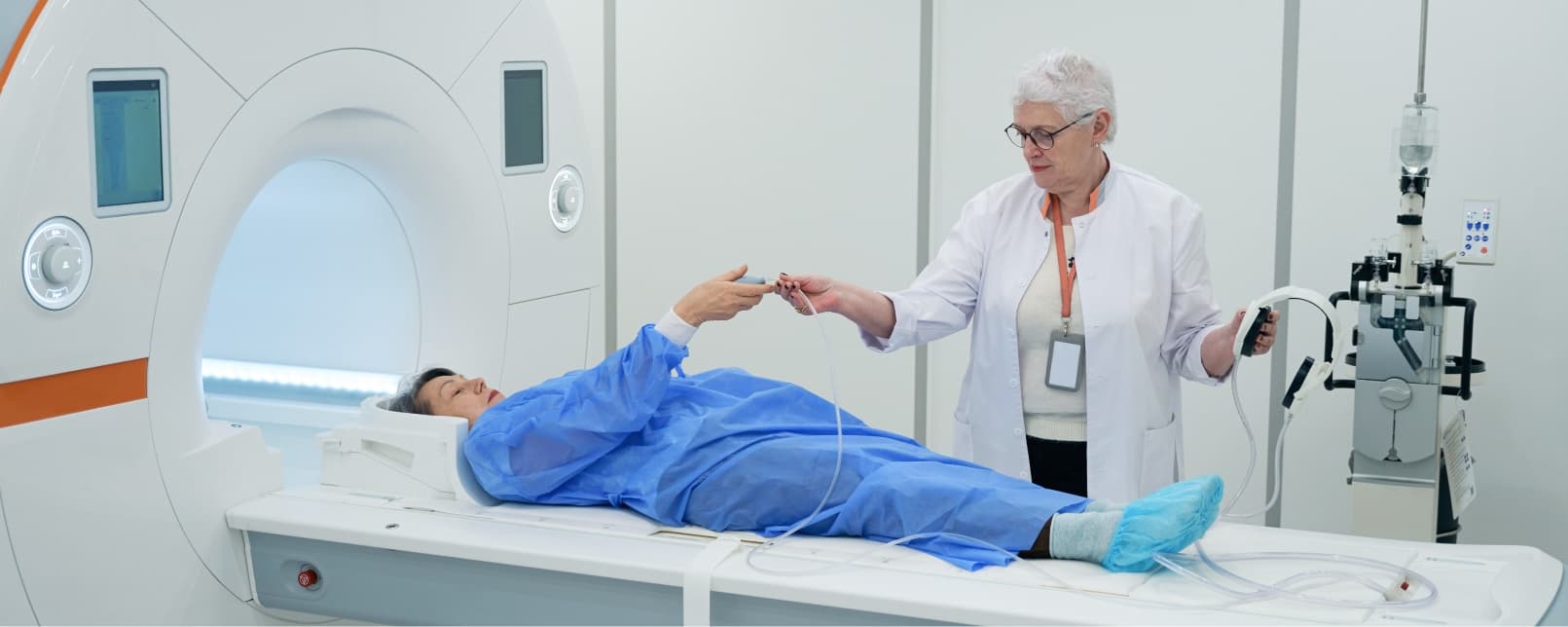
Precise diagnosis using advanced imaging & evaluation
20,000+
Successful Surgeries
When It's Needed
Conditions & Indications
When is this procedure recommended? These are the conditions where Hip Replacement Surgery typically offers the best outcome.
Experiencing these symptoms?
An accurate diagnosis is the first step. Get an expert evaluation from Dr. Hemendra Agrawal.
Book a ConsultationOsteoarthritis of the hip: the most common reason, causing progressive cartilage destruction and bone-on-bone contact
Avascular necrosis (AVN) of the femoral head, bone death due to disrupted blood supply, often from steroid use, alcohol, or trauma
Rheumatoid arthritis causing chronic inflammation and progressive hip joint destruction
Hip fractures in elderly patients (femoral neck fractures), requiring hemiarthroplasty or total hip replacement
Post-traumatic arthritis following acetabular fractures, hip dislocations, or femoral head fractures
Ankylosing spondylitis with hip joint involvement causing progressive stiffness and fusion
Developmental dysplasia of the hip (DDH) with secondary arthritis in adults
Failed previous hip surgeries including osteotomy, internal fixation, or prior hip replacement
Step by Step
How is Hip Replacement Surgery Performed?
A detailed walkthrough of the surgical process
Step 01 / 05
Pre-Operative Assessment
Complete medical evaluation, hip and pelvic X-rays (AP pelvis, lateral hip), MRI for complex cases, CT scan for 3D planning in dysplastic or revision cases. Templating for implant sizing. Medical optimization of diabetes, hypertension, and cardiac conditions. Pre-operative physiotherapy to strengthen hip abductors and improve walking ability.
Step 02 / 05
Surgical Approach: Direct Anterior Approach
Through a 10-12 cm incision over the front of the hip, the joint is accessed through the interval between the sartorius/rectus femoris and tensor fascia latae muscles, no muscles are cut or detached. This inter-nervous, inter-muscular approach preserves the hip abductors and external rotators, providing inherent stability against dislocation.
Step 03 / 05
Acetabular (Socket) Preparation
The damaged cartilage and bone of the acetabulum are removed using hemispherical reamers of progressively increasing size. The acetabular component (cup) is press-fit into the prepared socket at the optimal angle, approximately 40-45 degrees of inclination and 15-20 degrees of anteversion. Screws may be added for supplementary fixation. A ceramic or polyethylene liner is inserted into the cup.
Step 04 / 05
Femoral (Ball) Preparation
The femoral neck is cut at a precise level, and the femoral head is removed. The femoral canal is prepared with broaches of increasing size to accommodate the femoral stem. A trial stem and head are placed to check leg length, offset, stability, and range of motion. The final femoral stem (titanium, uncemented) is impacted into the canal, and a ceramic or metal femoral head is placed on the taper.
Step 05 / 05
Joint Reduction & Closure
The hip joint is reduced (ball placed into socket) and tested for stability in flexion, extension, internal rotation, and external rotation. Range of motion and leg length are verified. The incision is closed in layers. With the anterior approach, no precautions against posterior dislocation are needed, allowing earlier mobilization and fewer movement restrictions.
Facilities & Care
Advanced Surgery & Personalized Care
From a state-of-the-art operation theatre to attentive post-operative recovery: you're in expert hands at every step.
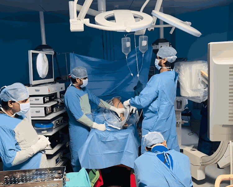
Modern Operation Theatre
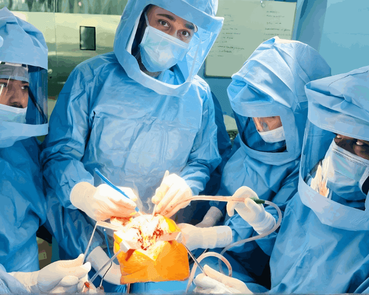
Precision Surgery in Progress
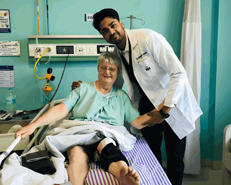
Attentive Post-Operative Care

Personalized Patient Support
The Outcome
Key Benefits
How this procedure transforms your life
Dramatic pain relief: 97% of patients report significant or complete pain relief after surgery
Restored hip mobility: sitting, walking, climbing stairs, putting on shoes, and crossing legs
Correction of leg length discrepancy (if present) for balanced, comfortable walking
Anterior approach: muscle-sparing technique with lower dislocation risk (0.5% vs. 3-5% with posterior approach)
Advanced bearing surfaces (ceramic-on-ceramic), virtually wear-free, ideal for younger active patients
Uncemented implants with biological fixation: designed to last 30+ years in appropriate patients
Earlier return to activities: many patients walk without support within 2-4 weeks
Improved overall health: increased physical activity improves cardiovascular fitness, mood, and bone density
Your Journey
Recovery Timeline
What to expect during your recovery journey, step by step
Immediate Post-Op
Walking with walker within 6-24 hours. With anterior approach, no hip precautions needed. Ankle pumps, quadriceps sets, and gluteal squeezes initiated.
Hospital Recovery
Independent walking with walker, stair climbing, bathroom independence. Discharge typically on day 2-3 with anterior approach. Home exercise program and physiotherapy referral provided.
Early Home Recovery
Walker to cane transition by week 2-3. Driving at 2-4 weeks. Daily exercises for hip strengthening. Outpatient physiotherapy twice weekly. Suture removal at 2 weeks.
Strengthening Phase
Walking without aids, return to desk work, light recreational activities. Swimming and cycling permitted. Progressive hip abductor and extensor strengthening.
Full Recovery
Return to all normal activities. Low-impact sports: golf, cycling, swimming, hiking. Avoid high-impact activities. Follow-up with X-rays at 6 months.
Insurance & Panels
Insurance & Cashless Options for Hip Replacement
Government panels and health insurance we help you claim, with cashless coordination and complete paperwork support.
CGHS
For central government employees & pensioners
ECHS
For ex-servicemen (defence) & dependents
RGHS
For Rajasthan government employees & pensioners
Railway Panel
For railway employees & pensioners
Ayushman Bharat
Cashless cover up to ₹5 lakh per family / year
MAA / Chiranjeevi
Rajasthan cashless cover · via Jan Aadhaar
Cashless Insurance
Cashless orthopedic surgery via your health insurer
Explore More
Other Services
Explore our full range of related procedures






FAQ
Frequently Asked Questions
Get answers to common questions about this procedure
Ready to Take the First Step?
Book a consultation with Dr. Hemendra Agrawal to discuss your condition and explore the best treatment options for you.