Partial Knee Replacement Surgery in Jaipur
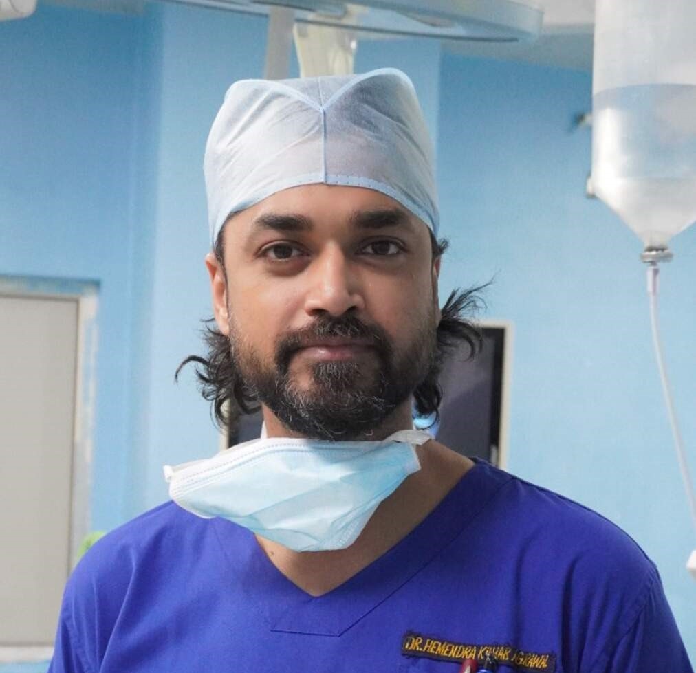
Unicompartmental Knee Arthroplasty by Dr. Hemendra Agrawal
What is Partial Knee Replacement?
Partial Knee Replacement, also known as Unicompartmental Knee Arthroplasty (UKA), is a bone-conserving surgical procedure that replaces only the damaged compartment of the knee joint while preserving the healthy compartments, intact ligaments (including the cruciate ligaments), and undamaged cartilage. Unlike total knee replacement which resurfaces all three compartments, partial knee replacement targets only the diseased area — providing a more conservative, tissue-preserving solution.
The knee joint has three compartments: the medial (inner) compartment, the lateral (outer) compartment, and the patellofemoral (front) compartment. In many patients, arthritis affects only one of these compartments. For these patients, partial knee replacement offers a less invasive alternative that preserves the natural biomechanics of the knee, resulting in a more natural-feeling joint with better proprioception (joint position sense) compared to total knee replacement.
Dr. Hemendra Agrawal is one of the few surgeons in Jaipur with extensive experience in partial knee replacement using robotic-assisted and computer-navigated techniques. These advanced technologies enable precise implant positioning that is critical for the success of unicompartmental replacement — even small alignment errors can lead to premature failure in partial replacements.
The minimally invasive approach used for partial knee replacement involves a smaller incision (7-10 cm), less bone removal, preservation of both cruciate ligaments and the unaffected compartment, reduced blood loss, less post-operative pain, faster rehabilitation, and quicker return to normal activities compared to total knee replacement.
Conditions & Indications
When is this procedure recommended?
Isolated medial compartment osteoarthritis (the most common indication — affecting the inner side of the knee)
Isolated lateral compartment osteoarthritis (affecting the outer side of the knee)
Avascular necrosis limited to a single femoral condyle
Post-traumatic arthritis confined to one compartment following a localized injury
Intact or functional anterior cruciate ligament (ACL) and posterior cruciate ligament (PCL)
Correctable varus or valgus deformity less than 15 degrees
Knee flexion range of at least 90 degrees with minimal flexion contracture (less than 10 degrees)
BMI preferably less than 35 (though not an absolute contraindication)
How is Partial Knee Replacement Performed?
A detailed walkthrough of the surgical process
Patient Selection & Assessment
Careful patient selection is the most critical factor for success. Dr. Agrawal performs a thorough clinical examination assessing ligament integrity, deformity correctability, and range of motion. Weight-bearing X-rays (AP, lateral, skyline, and stress views) and MRI confirm isolated compartmental disease with intact cruciate ligaments and preserved cartilage in the other compartments.
Robotic-Assisted Surgical Planning
A pre-operative CT scan is obtained to create a 3D model of the knee. The robotic system software generates a patient-specific surgical plan optimizing implant size, positioning, and alignment. The surgeon reviews and fine-tunes this plan before surgery, ensuring millimeter-level precision.
Minimally Invasive Approach
Through a small 7-10 cm incision, the affected compartment is accessed without disturbing the healthy compartments. The medial parapatellar approach allows visualization of the damaged compartment while preserving the cruciate ligaments, meniscus in the healthy compartment, and the quadriceps mechanism.
Precision Bone Preparation
Using robotic-assisted cutting tools with haptic boundary control, only the diseased bone and cartilage are removed — preserving maximum healthy bone stock. The femoral condyle and tibial plateau in the affected compartment are precisely prepared to receive the implant components. The robotic system prevents any cutting beyond the planned boundaries.
Implant Placement & Closure
The femoral and tibial components are cemented in place with the polyethylene bearing surface inserted. Limb alignment, ligament balance, and range of motion are verified. The incision is closed in layers. Due to the smaller incision and less tissue dissection, blood loss is typically less than 100 ml, and most patients do not require a drain.
Key Benefits
How this procedure transforms your life
Smaller incision (7-10 cm vs. 15-20 cm) — less tissue trauma and better cosmetic result
Preservation of both cruciate ligaments — resulting in more natural knee kinematics and proprioception
Faster recovery — most patients walk without aids within 3-4 weeks vs. 6-8 weeks for TKR
Less post-operative pain and reduced need for pain medications
Shorter hospital stay — typically 1-3 days vs. 3-5 days for TKR
Greater range of motion — typically 130-140 degrees flexion vs. 110-120 degrees for TKR
Preservation of healthy bone stock — allows conversion to total knee replacement if needed in the future
More natural-feeling knee — 90% of patients forget they have an artificial joint
Recovery Timeline
What to expect during your recovery journey
Immediate Post-Op
Standing and walking with walker within 6-12 hours. Less pain compared to TKR due to smaller incision and preserved tissues. Early range of motion exercises initiated.
Hospital Stay
Independent walking with walker, stair climbing, achieving 90+ degrees flexion. Most patients discharged by day 2-3. Home exercise program provided.
Early Recovery
Transition from walker to cane by week 2. Driving may resume at 2-3 weeks. Outpatient physiotherapy 2 times per week. Light household activities.
Active Recovery
Walking without aids, return to desk work, light recreational activities. Achieve 120+ degrees flexion. Swimming and cycling permitted.
Full Recovery
Return to most normal activities including gardening, golf, walking, and low-impact sports. Final assessment with X-rays at 3 months.
Other Services
Explore our full range of joint replacement procedures
Frequently Asked Questions
Get answers to common questions about this procedure
Ready to Take the First Step?
Book a consultation with Dr. Hemendra Agrawal to discuss your condition and explore the best treatment options for you.